


|
|

 Medical thermography which is also called Infrared or DII (Digital Infrared Imaging) is the process of obtaining highly detailed and sensitive infrared images of the human The FDA listed Breast Thermography as approved for breast cancer risk assessment in 1982. The Flir Model A infrared camera that we use at our center received it's 510(K) classification from the FDA in 2004. The FDA has not approved of Infrared as a "diagnostic" procedure as a stand-alone test since it is to be used adjunctively with other imaging techniques and other testing ordered by your physician. Medical thermography which is also called Infrared or DII (Digital Infrared Imaging) is the process of obtaining highly detailed and sensitive infrared images of the human The FDA listed Breast Thermography as approved for breast cancer risk assessment in 1982. The Flir Model A infrared camera that we use at our center received it's 510(K) classification from the FDA in 2004. The FDA has not approved of Infrared as a "diagnostic" procedure as a stand-alone test since it is to be used adjunctively with other imaging techniques and other testing ordered by your physician.
Modern Infrared technology benefited and evolved rapidly following millions of dollars of military money being spent on the surface to air missile program during the Gulf War under General Secord and more recently with "Smart Infrared Helmets" on medics in the Iraq War. The Flir Model A camera that is used in our clinic had also been used for military applications for years.
Breast cancer concerns have been mounting and self-examinations, clinical examinations by your doctor and mammogram's have been widely promoted as means of saving lives from breast cancer. However, the mortality of breast cancer is essentially unchanged in the past 65 years because none of these techniques provide true early detection. One factor is that Mammography detects tissue changes that indicate disease is already present. A Thermography screening can detect metabolic tissue changes that if left untreated may later present as disease. Abnormal Thermology can be an indication of risk. Thermology also has an advantage for all women with dense fibrocystic breasts and is recommended for women of all ages. What the "Proactive Breast WellnessTM program" teaching module is addressing are ways for women to try to make changes, that will alter in a positive way, the initial abnormal test values. This is a five CD set that is about to be available nationally as well as through our web site. For more information, please view the Proactive section on this site. One of the goals of the "Proactive Breast WellnessTM program" is to help support the immune system so the inflammation or cellular activity does not continue to get worse.
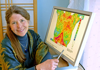
 Thermology is a passive type of testing. This means thermography does NOT use any form of ionizing radiation (such as does X-ray) as it evaluates the features of activity and operations of the body. The equipment we use, which is a Flir Model A40 infrared thermography camera makes tens-of-thousands of detailed measurements of skin temperature and the camera is placed about 4 to 8 feet from your body and there is no compression and it is 100% safe. This camera is accurate to within 0.06 degrees Celsius and it runs at video speed. One image can be enlarged into an 8 by 11 inch print and can easily be sent via jpeg to a surgeon or radiologist. Our software can be changed into 12 different color palettes including a gray inverted palette to visualize larger vessels that may be feeding a tumor which will be further evaluated by other structural studies. You are also able to evaluate the effect of hormone replacement, the effects of not eating organically grown or raised foods that have excessive bovine growth hormones. Infrared is able to visually detect estrogenic effects in the breast that may be related to the risks of being toxically exposed to estrogen mimickers as found in herbicides /pesticides and other chemicals. (Please go to the "Proactive Breast Wellness" section of this site to view our ground breaking research on a power point presentation that demonstrates what herbicides and pesticides are doing to breast tissue). Thermology is a passive type of testing. This means thermography does NOT use any form of ionizing radiation (such as does X-ray) as it evaluates the features of activity and operations of the body. The equipment we use, which is a Flir Model A40 infrared thermography camera makes tens-of-thousands of detailed measurements of skin temperature and the camera is placed about 4 to 8 feet from your body and there is no compression and it is 100% safe. This camera is accurate to within 0.06 degrees Celsius and it runs at video speed. One image can be enlarged into an 8 by 11 inch print and can easily be sent via jpeg to a surgeon or radiologist. Our software can be changed into 12 different color palettes including a gray inverted palette to visualize larger vessels that may be feeding a tumor which will be further evaluated by other structural studies. You are also able to evaluate the effect of hormone replacement, the effects of not eating organically grown or raised foods that have excessive bovine growth hormones. Infrared is able to visually detect estrogenic effects in the breast that may be related to the risks of being toxically exposed to estrogen mimickers as found in herbicides /pesticides and other chemicals. (Please go to the "Proactive Breast Wellness" section of this site to view our ground breaking research on a power point presentation that demonstrates what herbicides and pesticides are doing to breast tissue).
There are no temperature probes that some of the older technology used over 13 years ago to capture temperature data from the skin. Simply touching the skin with a probe will cause the client to react and usually vaso-constrict or pull away by the act of being touched. This will of course alter the readings hence not making the probe technology very accurate. The images produced from our camera are really electronic data of absolute temperature measurements that can be viewed as an electronic image presenting a spectrum of colors that indicate increased or decreased levels of infrared radiation (heat) being emitted from you own body's surface. Cancers at different stages can have an increased tissue metabolism resulting from rapid multiplication of the cells. Increased metabolism can generate heat that may be detected as an asymmetry in your scan. Thermology detects the resulting heat from biochemical reactions and physiology and is distinctly different from tissue structure-based diagnostic methods, such as MRI, mammogram's, and ultrasounds. Thermology does not replace these other diagnostic methods but rather; it adds to the other structural testing to improve their diagnostic value and to complement your comprehensive program of breast evaluation. Breast thermology is particularly effective in instances where mammography is compromised; such as in women before menopause, women who have used hormone replacement therapy (HRT), have glandular or dense breasts, have fibrocystic disease, had prior biopsies, have implants or reconstructive/ reduction surgery with a lot of scar tissue, are pregnant, are nursing or have small or large breasts, or have had toxic exposures of herbicides/ pesticides/xenoestrogens/estrogen disruptors.
Breast thermography has a very high, on average 90% sensitivity, identifying the specific tissue thermal signals that act as risk markers and changes associated with breast cancer. There is only a 10% false positive rate in Infrared especially on the initial studies of an individual. This is why a baseline and subsequent periodic imaging has been beneficial to closely observe any changes over time. This allows the practitioner to encourage the woman to go for further structural studies which may include an MRI, to rule out any detectable disease. A questionable/ borderline TH3 thermology feature may resolve in about 70% of the women in who are aggressive with the "Proactive Breast WellnessTM program" protocol when they work on balancing their hormones, decrease their estrogen dominance, make lifestyle and dietary changes. This is very encouraging and the educational program has been empowering women to improve their health in many areas.
A recent study published in October of 2008 in the American Journal of Surgery, and performed at Cornell Medical School, found thermography to be 97% sensitive. Infrared had identified 58 of the 60 tumors in women with known cancers. They were using the same camera we have at our center. Read Full Article
There was a large study done on 58,000 women with breast complaints that was written up in "Cancer" in 1980, Vol. 56,45-51. Of that 58,000 group of women, 1,245 patients had a borderline TH3 TH4 or TH5 infrared thermograms but had normal mammograms, ultrasounds, physical exam and biopsy. Thirty-eight percent of the women with normal breasts and 44% of those with mastopathy developed biopsy proven breast cancer within five years!!! Since the modern infrared is now being able to detect asymmetrical temperatures and patterns determined as outside normal physiological variations, these asymmetries can be monitored and combined with other modes of evaluation to increase the odds of early detection. This makes Infrared Thermography the Best Risk Assessment Tool we now have available! The infrared images are seeing physiological activity where groups of cells are now clustering possibly prior to lump formation at about 2mm or the thickness of a credit card. At a certain growth point, the abnormal cells will require more circulation to the area to provide the growing mass of cells with more nutrition. The development of these new blood vessels (angiogenesis) of a solid malignant tumor must occur when it has grown too large for simple diffusion from existing vessels to provide for the metabolic needs of the cells of the tumor. The process of angiogenesis begins when a malignant tumor is about 150 micrometers (0.15mm) in diameter and must be extensively developed by the time a tumor is 1-2 mm in diameter. Angiogenic blood vessels are unstable and do not have the ordered structure of normal blood vessels. In fact, angiogenic vessels are of a primitive structure without any connection to the autonomic nervous system and no vascular smooth muscle content. Infrared is able to show circulation changes and the patterns are frequently chaotic in nature. This is a good indicator to send a woman in for further structural studies. False-negative errors are rare and usually a consequence of a latent (resting, non-active) stage in the development of breast cancer. Since breast cancers take about 10-12 years to grow to a stage that can be felt, the hope is that these women will have the issue identified earlier so they can come in for treatment earlier and hence increasing their survival rate. Imaging centers that offer mammography with infrared and scanning ultrasound, the combined sensitivity has approached a 98% catch rate. Diagnosing breast cancer early is of critical importance to surviving the disease. The survival of stage one disease in approximately ninety-one percent while survival of stage three drops to approximately forty percent (40%). Since Infrared thermography is the best and earliest risk detection tool for metabolic changes available to modern medicine, we are hoping that this technology will be in more imaging centers in the United States soon.
To learn more about thermography, please visit our interpreter’s site by Dr.
William Amalu for a more in depth look at this area. Click Here.
|
Home | Physician's
Area | Proactive Breast WellnessTM program |
What Is Thermography
Cost/FAQ
|
Research | About Us
| Store |
Publicity/Testimonials | Contact
Last Update:
This web site is protected by copyright. No part of this web site may be reproduced in any form or by
any means, including computer printouts, photocopying of printouts, or utilized by any information
storage and retrieval system without written permission from the copyright owner. Computer printouts
for personal use, and not dissemination, are permitted.
Legal Disclaimer |
web site design by
M. England
|